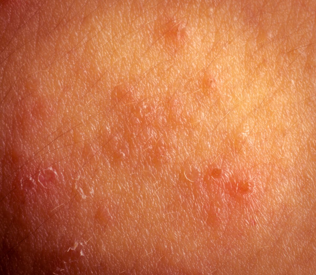
“Atopic dermatitis (AD), commonly referred to as eczema, is a prevalent condition in paediatric populations worldwide. It presents variably across different age groups and skin types, posing diagnostic and management challenges for healthcare professionals. This blog provides an overview of the presentation of AD in children of different ages, with a particular focus on variations in skin colour.”
Neonates and Infants (0-2 years)
In neonates and infants, AD often presents as erythematous, scaly, and crusted plaques on the face, scalp, and extensor surfaces. The nappy area is typically spared due to the moisture barrier provided by nappies. Pruritus is a key feature and may lead to sleep disturbances. In infants with darker skin, the erythema may not be as apparent, and the skin may appear brown, purple, grey or ashen. Post-inflammatory hyperpigmentation or hypopigmentation may also occur, leading to patches of skin that are darker or lighter than the surrounding skin.
Toddlers (2-5 years)
In toddlers, AD typically localises to the flexural folds of the elbows and knees, wrists, ankles, and hands. Periorbital and perioral involvement is also common. Lichenification, papulation, and hypopigmentation or hyperpigmentation may be observed due to chronic inflammation and scratching. In children with darker skin, the lichenification may appear greyish or dark brown, and there may be prominent follicular accentuation.
School-age children (5-12 years)
In school-age children, AD continues to favour flexural areas such as antecubital and popliteal fossae. The skin may appear lichenified and xerotic, with accentuated skin markings. Secondary bacterial infections due to Staphylococcus aureus can occur due to persistent scratching. In children with darker skin, the xerosis may be more pronounced and appear ashy, and post-inflammatory hyperpigmentation or hypopigmentation can lead to significant changes in skin colour.
Adolescents (12+ years)
In adolescents, AD may persist in flexural areas, but also commonly affects the hands and feet. The skin may exhibit xerosis and lichenification. Nummular eczema and pityriasis alba are common concurrent conditions. In adolescents with darker skin, the nummular eczema may appear as well-defined, round or oval patches that are darker or lighter than the surrounding skin, and the pityriasis alba may present as hypopigmented patches on the face.
It’s crucial to remember that AD is a clinical diagnosis with variable presentations. It’s influenced by numerous factors, including genetic predisposition, environmental triggers, and the patient’s immune status. Management involves a multifaceted approach, including regular emollient use, trigger avoidance, and topical corticosteroids or calcineurin inhibitors for flare-ups. Always consider referral to dermatology for severe or refractory cases. Remember, while AD can be a challenging condition to manage, many children experience a decrease in symptoms or remission during adolescence. With the right care and management, AD can be effectively controlled.
Looking to enhance your understanding of paediatric eczema management? Check out the article ‘Understanding and Applying Emollients in Paediatric Eczema’ on Practitioner Development UK Ltd’s website. This guide provides valuable insights into the best practices for applying emollients to children with eczema.
References
- Stanford Medicine Children’s Health, no date. Atopic Dermatitis in Children. Available at: https://www.stanfordchildrens.org/en/topic/default?id=atopic-dermatitis-eczema-in-children (Accessed: 29 January 2024).
- National Eczema Association, no date. Eczema on black skin: pictures, symptoms and treatment. Available at: https://nationaleczema.org/eczema-on-black-skin/ (Accessed: 29 January 2024).
- Cleveland Clinic, no date. Atopic Dermatitis: What It Is, Symptoms, Causes & Treatment. Available at: https://my.clevelandclinic.org/health/diseases/9998-atopic-dermatitis-eczema (Accessed: 29 January 2024).


