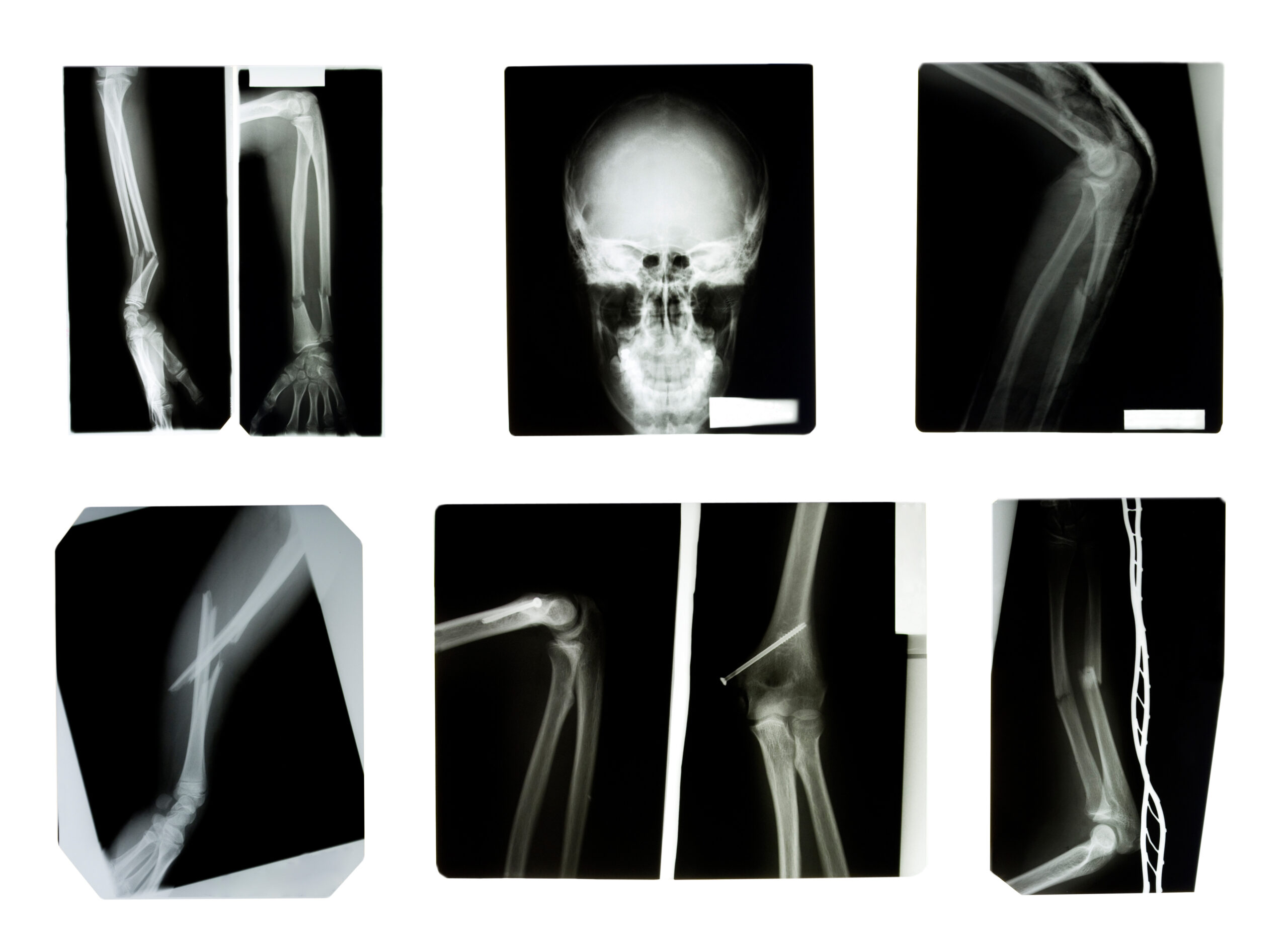
Interpreting an X-ray is a fundamental skill in the medical field, particularly for healthcare professionals working in primary care settings. X-rays, a form of electromagnetic radiation, can penetrate human tissue, producing images that can reveal signs of disease or injury. These images are invaluable tools for diagnosing a wide range of conditions.
In procedures such as radiography, computed tomography, and fluoroscopy, X-rays provide still images or allow for the observation of motion within the body. When it comes to diagnosing musculoskeletal injuries, X-rays are often the first line of investigation.
However, interpreting an X-ray requires a systematic approach. One commonly used method is the ABC principle:
A – Alignment and Adequacy: The alignment refers to the positioning of the bones in relation to each other. Any misalignment could indicate a dislocation or fracture. Adequacy refers to the quality of the X-ray image. An adequate X-ray ensures that the beam penetration is neither over nor underexposed and includes the joints above and below the area of concern.
B – Bones: Outline and Density: The outline of each bone should be smooth and continuous. Any interruption could suggest a fracture. The density of each bone should be assessed to identify whether it is radio-lucent (indicating thinner bone, as in osteoporosis) or radio-opaque (thicker than surrounding bones, as seen in conditions like chronic osteomyelitis, Paget’s disease, osteochondritis).
C – Cartilage: Outline, Joint Space, and Loose Bodies: The condition of the cartilage should be assessed. Any signs of wear, damage, or evidence of cartilage fragmentation should be noted. Changes in joint space width can indicate cartilage loss or joint effusion.
In addition to the ABC principle, it’s also important to consider the patient’s history, symptoms, and physical examination findings when interpreting an X-ray. This comprehensive approach can help ensure a more accurate diagnosis.
For healthcare professionals looking to further their understanding of X-ray interpretation, the article ‘The Red Dot System in X-Ray Interpretation: Development and Effectiveness’ on the PDUK website is a valuable resource. It provides a look at the Red Dot system, a method used by radiographers to indicate areas of concern on an X-ray that require further review by a radiologist. This system has been found to be effective in improving the accuracy of initial X-ray interpretations, particularly in busy clinical settings where immediate radiologist review may not be possible.
Whether you’re a seasoned professional looking to refresh your knowledge or a student seeking to learn more about X-ray interpretation, this article is a valuable resource. Don’t miss out on this opportunity to expand your expertise!
References
1.BMC Emergency Medicine (2023) ‘Radiographer-led discharge for emergency care patients, requiring projection radiography of minor musculoskeletal injuries: a scoping review’, BMC Emergency Medicine, available at BMC Emergency Medicine.
- Radiopaedia (2023) ‘Investigation of ankle injury (summary)’, Radiopaedia, available at Radiopaedia.
- The Royal College of Radiologists (2023) ‘Standards for interpretation and reporting of imaging investigations’, The Royal College of Radiologists, available at [The Royal College of Radiologists].


